Prof. N. Reha Tandoğan, M.D. - Asım Kayaalp, M.D.
What is a meniscal root tear?
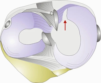
Meniscal root tears are tears within 1 cm of the bone insertions of the meniscus. (Figure 1) The importance of meniscus root tears has recently been elucidated, and treatment options have changed dramatically in recent years. Root tears are important because if left untreated, the ability of the meniscus to bear and transfer load is lost; leading to early cartilage damage and arthritis. Experimental studies have shown that repairing the root tear restores the normal biomechanics of the knee joint and prevents osteoarthritis
Root tears are not the same for the medial and lateral meniscus
Root tears of the lateral(outer) and medial (inner) meniscus are completely different. Lateral meniscal root tears are seen in younger patients and are almost always associated with an injury to the anterior cruciate ligament (ACL), one of the most common knee injuries in sports like soccer, basketball and skiing. They are difficult to visualize on magnetic resonance imaging and may sometimes be discovered during surgery for ACL reconstruction. Repair of these tears is essential to prevent progressive cartilage damage and increased loads on the reconstructed ACL, leading to increased risk of failure of ligament surgery.Medial meniscal root tears are usually seen in middle aged and elderly patients and are a part of the degenerative process of the knee. There is usually no history of major trauma. Sometimes a painful pop may be noticed while kneeling. Obesity is an important risk factor. Once torn, the meniscus is pushed out of the joint during weightbearing, leading to a total loss of meniscal function. Medial root tears are also associated with spontaneous osteonecrosis of the knee, starting with bone marrow edema followed by localized bone death and collapse of the condyle. MRI diagnosis is easier on the medial side with the typical ghost sign and extrusion of the meniscus. We are one of the first groups to describe this entity in 2008 (Ozkoc G, Circi E, Gonc U, Irgit K, Pourbagher A, Tandogan RN. Radial tears in the root of the posterior horn of the medial meniscus. Knee Surg Sports Traumatol Arthrosc. 2008 Sep;16(9):849-54.)
How is a root tear diagnosed?
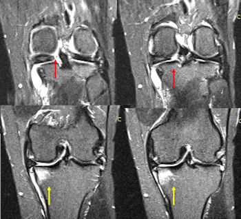
The diagnosis of a root tear is made by careful history and clinical examination followed by imaging techniques. Some lateral root tears can only be diagnosed during arthroscopy. Magnetic resonance imaging (MRI) is the most accurate imaging technique to diagnose a root tear (Figure 2). Your doctor may also require some X-rays to evaluate the degree of osteoarthritis and limb alignment.
What are the treatment options for meniscal root tears?
Lateral meniscal root tears should always be repaired during your ACL surgery. Specific instruments have been developed to make this repair easier. (Video 1) Lateral root tears have a high healing rate.Video
In contrast, medial root tears have a variety of treatment options and your doctor will choose the most suitable one for you. Root tears may become asymptomatic in elderly patients with rest, limitation of weight-bearing using a cane/crutches and anti-inflammatory medication. However, the progression of osteoarthritis is inevitable. This option is preferred when you already have arthritis in the joint and are not a surgical candidate. Removal of the tear with knee arthroscopy may provide short term pain relief but symptoms may get worse with progression of osteoarthritis. These two were the only options until the last 5 years. However, the importance of these tears were recognized in recent years and techniques to preserve the meniscus were developed.
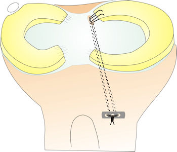
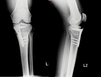
The most effective treatment of medial root tears is arthroscopic repair (Figure 3). This option is preferable in middle aged patients without advanced cartilage damage. The healing rate is about 70%; and if successful, prevents the progression of arthritis. Root repair may be combined with bone procedures such as a high tibial osteotomy to correct the alignment of the limb and to unload the repair site (Figure 4).


If there is advanced cartilage damage or avascular necrosis of the femoral condyle in addition to the root tear, your surgeon might choose to perform a partial knee replacement. . The damaged joint surfaces are covered with metal and polyethylene implants, reconstructing the medial side of the knee. This is a very successful operation with over 90% good to excellent outcomes at 10 years.
